 Loading... Please wait...
Categories
 New Products
-
 $6,800.00
-
 $4,670.00
-
 $11,600.00
-
$6,600.00$5,480.00
-

$3,250.00$2,880.00
Our Newsletter
- Home
- Medical Equipment
- Carl Zeiss Cirrus HD-OCT 5000
- Home
- Ophthalmology
- Carl Zeiss Cirrus HD-OCT 5000
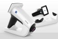
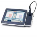
Carl Zeiss Cirrus HD-OCT 5000

Product Description
CARL ZEISS Cirrus HD-OCT 5000 provide highest resolution visualization capabilities and track retinal pigment epithelial integrity using Advanced RPE Analysis and assess glaucomatous loss with Ganglion Cell Analysis. Cirrus HD-OCT 5000 come with features an active eye-tracking mechanism that able to trace and compensate eye motion in real time and FastTrac retinal tracking to reduces eye motion artifacts without sacrificing patient
Cirrus OCT come with version 9.5 software can acquire 68,000 scans per second with fundus photos
Cirrus Review Software adds value in these critical areas:
- Allows detailed analysis of data in a doctors office, patient exam lane, or research center.
- Facilitates patient education by bringing dynamic display and 3D Advanced Visualization to a convenient location.
- Increases instrument availability for improved patient workflow.
- Supports simultaneous connection of up to ten review stations.
Cirrus Review provides dynamic viewing capabilities on a Windows based computer connected to a Cirrus 3.0 instrument database through a peer to peer office network. A single review software license allows up to 10 simultaneous review workstation connections.Minimum System Requirements
The system running Cirrus Review Software must meet the following minimum requirements:
- Operating systems: Windows XP Service Packs 2 and 3, Windows Vista Ultimate, Business and Enterprise supported. 64-bit operating systems are not supported.
- Processor: Intel Pentium IV, at least 1.5 GHz,or AMD Athlon, at least 1.8 GHz. Intel processors are recommended.
- Memory: 1 GB RAM; when running additional applications concurrently, more memory may be required.
- At least 20 GB available hard disk space.
- CD-ROM drive (for installation)
- 1024 x 768 pixel screen resolution; 32 bit color.
- Network interface card 100 Mbps
Note: Not all resolutions will work properly on wide-screen monitors.Cirrus analysis involves computational processing of large data sets,and therefore the review software performs best on a powerfulcomputing platform.
Specifications:
- OCT Imaging
- Methodology: Spectral domain OCT
- Optical source: Superluminescent diode (SLD), 840 nm
- Scan speed: 27K- 68K A-scans per second
- A-scan: 2.0 mm (in tissue), 1024
- Axial resolution: 5 μm (in tissue)
- Transverse resolution: 15 μm (in tissue)
Warranty Information

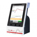
Customers Who Viewed This Product Also Viewed
-
$6,800.00$5,440.00
-
 $2,100.00
-
$3,400.00$2,920.00
-
 $13,700.00
Add to Wish List
Related Products
-
-

-
ConMed System 5000 Electrosurgical $2,100.00
-
-